

PROTECT YOURSELF with Orgo-Life® QUANTUM TECHNOLOGY
Orgo-Life the new way to the future Advertising by Adpathway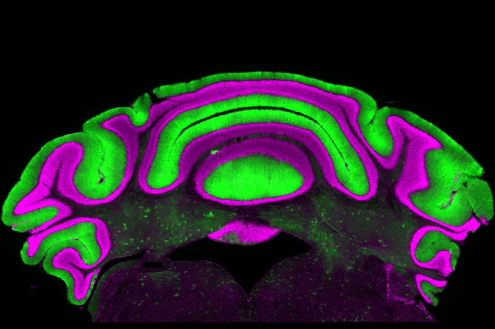 A new study from Meike van der Heijden’s lab at the Fralin Biomedical Research Institute at VTC challenges assumptions about brain activity in movement disorders involving Purkinje cells in the cerebellum, shown here in green, and deep nuclei cells, located in the dark areas with flecks of green in the center of this image. Credit: Meike van der Heijden/Virginia Tech
Scientists discovered that a key brain signal may have been pointing researchers in the wrong direction, potentially changing how movement disorders are studied and treated.
A new study from a Virginia Tech neuroscientist at the Fralin Biomedical Research Institute at VTC is prompting researchers to rethink a basic assumption about how chronic movement disorders are studied.
The research focuses on dystonia, ataxia, and tremor, three neurological disorders that cause involuntary movements including painful muscle contractions, abnormal postures, and shaking. Although these conditions have different symptoms, they all arise from problems in the cerebellum, the part of the brain responsible for coordinating movement.
For years, neuroscientists have relied on the activity of one type of brain cell to infer what another type is doing. The new findings suggest that approach may not provide an accurate picture.
Two Brain Cell Types May Not Be Closely Linked
Within the cerebellum, Purkinje cells normally suppress the activity of neurons in the deep cerebellar nuclei. Because of this direct connection, researchers have generally assumed that measuring Purkinje cell activity would reliably reflect what is happening in the deeper neurons.
A new study led by Meike van der Heijden, however, challenges that idea. Published in the Journal of Physiology, the research found that even though the two cell types are anatomically connected, the activity of one is a poor indicator of the activity of the other.
“We see that there’s not a clear linear relationship between activity in the Purkinje cells and in the deep nuclei cells. So there’s very limited predictive power in monitoring one to understand what’s going on in the other,” said Van der Heijden, assistant professor at the institute.
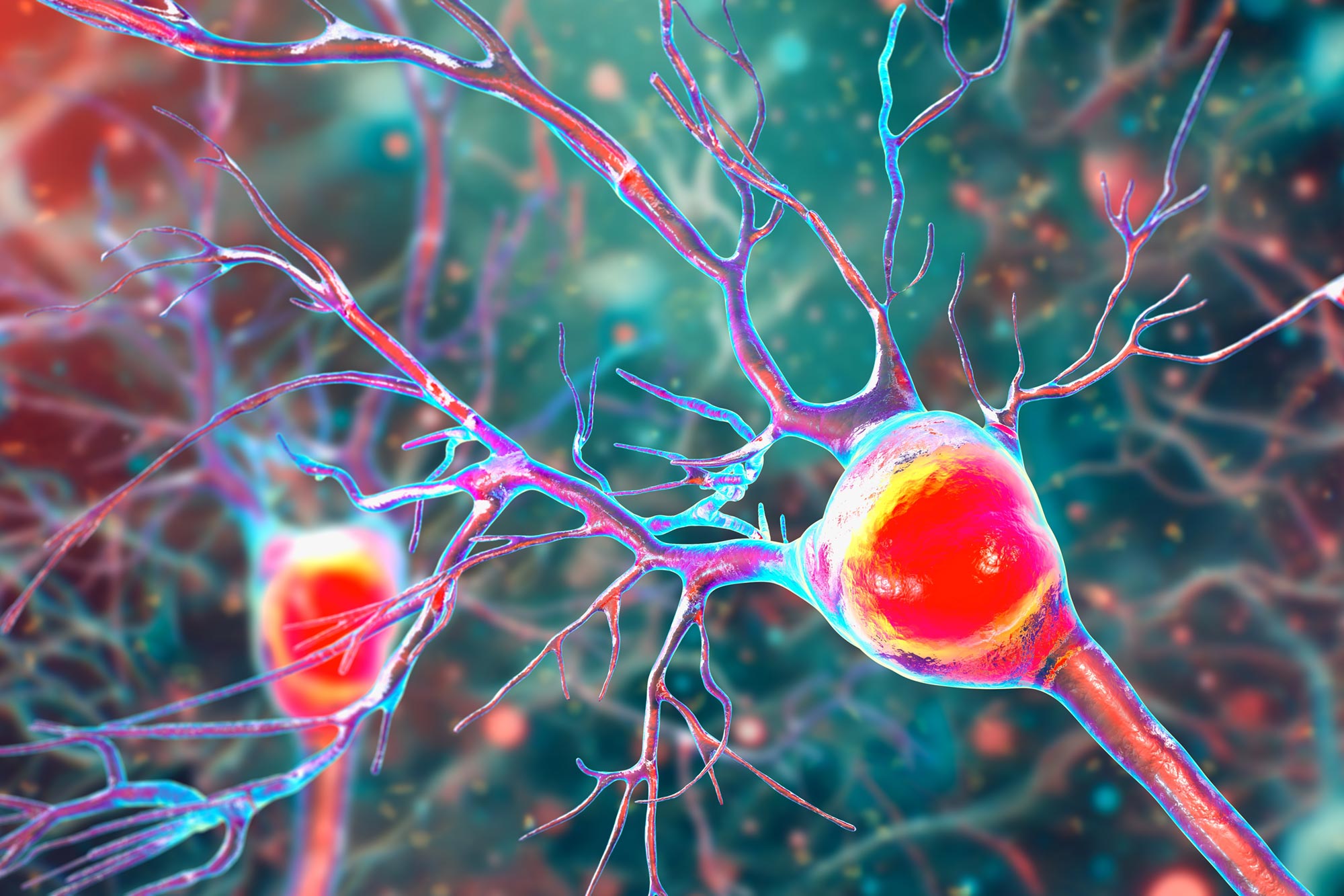 Purkinje cells are large neurons in the cerebellum that help coordinate movement, balance, and motor learning. They act as the brain’s primary control cells for fine-tuning movement by regulating signals sent to deeper brain circuits. Credit: Shutterstock
Findings Could Influence Future Treatments
The discovery has important implications for both research and the development of therapies for cerebellar movement disorders.
“Purkinje and cerebellar deep nuclei cell activity is disrupted in a disease state, and a better understanding of the relationship between these neuron types will ultimately help optimize treatments for diseases such as dystonia, ataxia, and tremor,” said Alyssa Lyon, a doctoral candidate in Virginia Tech’s Translational Biology, Medicine, and Health Graduate Program and the study’s first author.
Purkinje cells are located in the cerebellum’s outer layer, making them much easier to record than deep cerebellar nuclei cells, which lie farther beneath the brain’s surface. Because they are more accessible, Purkinje cells have long been treated as a convenient biomarker for the activity of the deeper neurons.
Under normal conditions, Purkinje cells inhibit deep cerebellar nuclei cells. As a result, researchers expected that higher Purkinje cell activity would correspond with lower activity in the deep nuclei, and vice versa.
Researchers Found No Meaningful Correlation
To test that assumption, the research team analyzed a database of electrophysiology recordings collected from pre-clinical models of cerebellar disease. Despite the expected relationship, they found no significant correlation between the activity of the two neuron types.
“We suggest that if you want to know how the cerebellum is behaving in a disease state, you have to look at the deep nuclei neurons, not just the Purkinje cells,” said Van der Heijden, who also holds an appointment in Virginia Tech’s School of Neuroscience.
She also cautioned that treatments designed to alter Purkinje cell activity should not automatically be expected to produce corresponding changes in the deep cerebellar nuclei.
“This is a cautionary tale for understanding cerebellar activity in disease, but also for treating these challenging diseases,” Van der Heijden said. “We need to be very careful in making assumptions, and to actually do experiments to test our hypotheses.”
Reference: “Steady-state Purkinje cell activity has limited predictive power for cerebellar output in disease” by Alyssa M Lyon, Viviana Hernandez-Castanon and Meike E van der Heijden, 20 April 2026, The Journal of Physiology.
DOI: 10.1113/JP290000
Never miss a breakthrough: Join the SciTechDaily newsletter.
Follow us on Google and Google News.






















 English (US) ·
  French (CA) ·
  French (FR) ·