
PROTECT YOURSELF with Orgo-Life® QUANTUM TECHNOLOGY
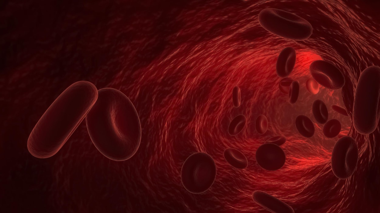
Orgo-Life the new way to the future Advertising by Adpathway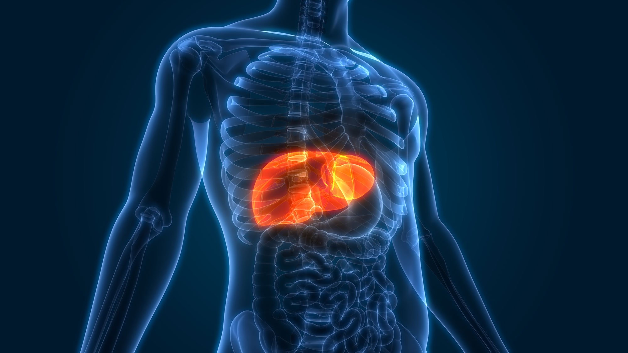 Scientists have created injectable “mini livers” that restored important liver functions in mice for weeks. Credit: Shutterstock
Engineered tissue grafts could help perform key liver functions and benefit thousands of people living with liver failure.
The liver is one of the body’s hardest-working organs, carrying out hundreds of vital jobs, from filtering toxins and metabolizing medications to producing proteins essential for blood clotting. Yet when it fails, the only definitive treatment is often a transplant, a solution limited by a chronic shortage of donor organs.
MIT engineers have now developed injectable “mini livers” that, in mice, survived for at least two months while performing many of the functions of healthy liver tissue.
“We think of these as satellite livers. If we could deliver these cells into the body, while leaving the sick organ in place, that would provide booster function,” says Sangeeta Bhatia, the John and Dorothy Wilson Professor of Health Sciences and Technology and of Electrical Engineering and Computer Science at MIT, and a member of MIT’s Koch Institute for Integrative Cancer Research and the Institute for Medical Engineering and Science (IMES).
Bhatia is the senior author of the study, which was published in the journal Cell Biomaterials. MIT postdoc Vardhman Kumar is the paper’s lead author.
Restoring liver function
The liver carries out about 500 vital jobs, from helping control blood clotting to clearing bacteria from the blood and breaking down drugs. Many of these tasks depend on hepatocytes, the liver’s main functional cells.
For more than a decade, Bhatia’s lab has been developing ways to restore hepatocyte activity without requiring a surgical liver transplant. One strategy is to place hepatocytes inside a biomaterial such as a hydrogel, but that approach still requires surgery to implant the gel.
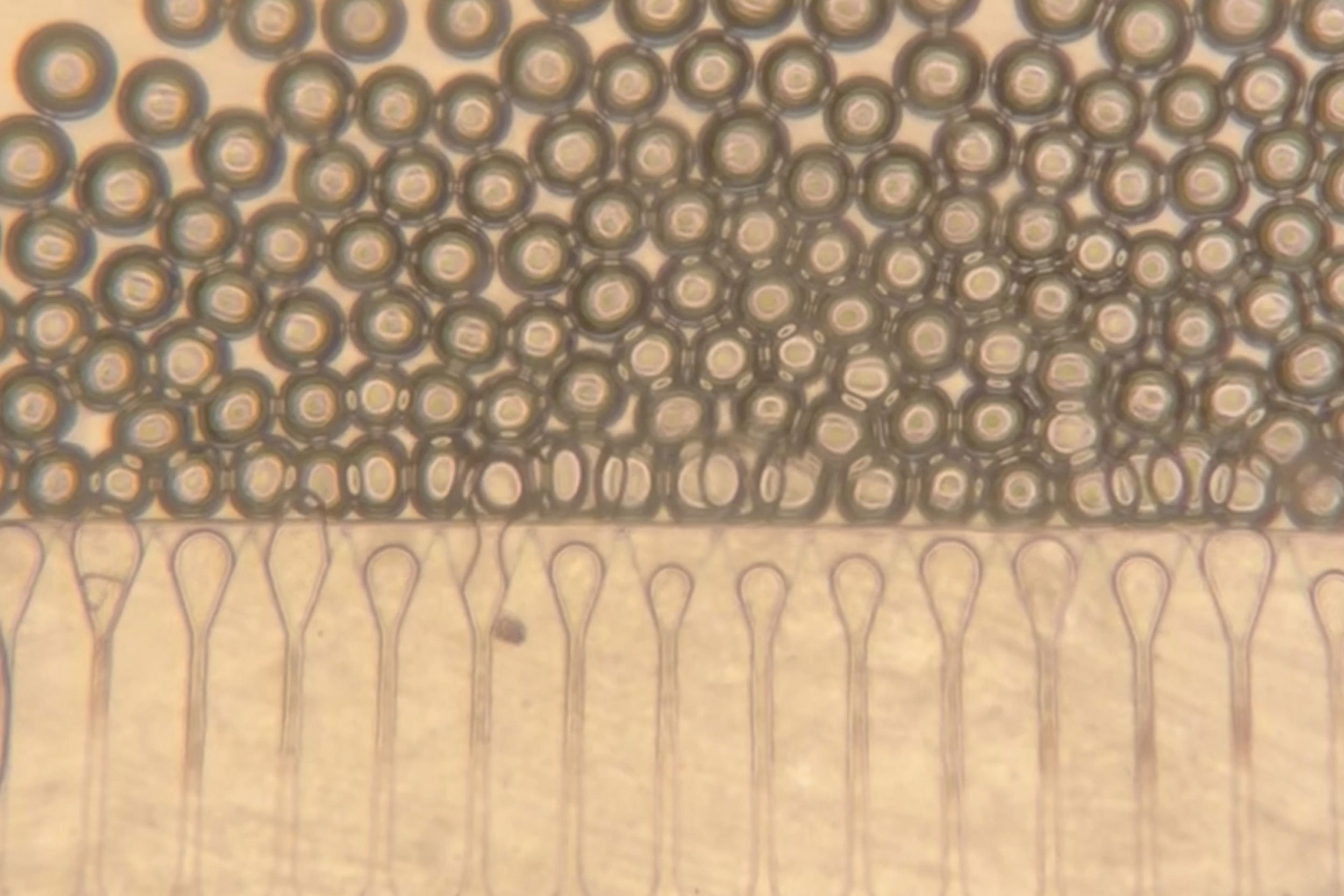 Researchers used a microfluidic device to generate hydrogel microspheres of uniform shape and size. These spheres are then mixed with hepatocytes and injected into the body, where they form stable mini livers. Credit: Bhatia Lab
Injecting hepatocytes directly into the body could avoid that surgery. In this study, Bhatia’s lab aimed to make that approach more effective by giving the cells an engineered environment that could help them survive and allow doctors to track graft health without another invasive procedure.
The solution was to inject the cells together with hydrogel microspheres. These tiny spheres help the cells remain clustered and connect with nearby blood vessels. They can behave like a liquid when packed together, which allows them to pass through a syringe, then return to a solid structure once inside the body.
In recent years, hydrogel microspheres have been studied as tools for wound healing because they allow cells to move into the spaces between the spheres and form new tissue. In the new work, the MIT group adapted the same basic idea to help hepatocytes build a stable graft after injection.
“What we did is use this technology to create an engineered niche for cell transplantation,” Kumar says. “If the cells are injected in the absence of these spheres, they would not integrate efficiently with the host, but these microspheres provide the hepatocytes with a niche where they can stay localized and become connected to the host circulation much faster.”
The injected material also contains fibroblast cells, which support hepatocyte survival and encourage blood vessels to grow into the graft.
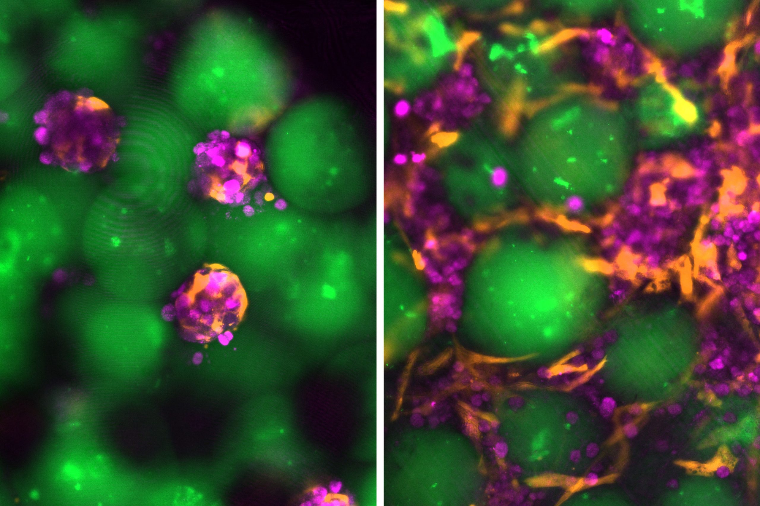 The microspheres (green), hepatocytes (magenta), and supporting fibroblasts (orange) assemble and reorganize into engineered liver grafts over time. Image shows a comparison of day zero (left) and day fourteen (right) of the grafts cultured in the laboratory. Credit: Bhatia Lab
Working with Nicole Henning, an ultrasound research specialist at the Koch Institute, Bhatia’s lab developed an ultrasound-guided syringe method to place the cell mixture in the body. The same imaging method can also be used after injection to follow the implant’s stability over time.
For this study, the mini livers were placed in fat tissue in the abdomen. Future versions could potentially be delivered to other parts of the body, including the spleen or areas near the kidneys. If the graft has enough room and a strong blood supply, the injected hepatocytes can act much like hepatocytes inside the liver.
“For a vast majority of liver disorders, the graft does not need to sit close to the liver,” Kumar says.
An alternative to transplantation
In mouse experiments, the mixture of liver cells and microspheres was injected into fatty tissue called perigonadal adipose tissue. After the cells settled in place, they formed a dense and stable structure. As time passed, new blood vessels grew into the graft, helping keep the hepatocytes alive and functional.
“The new blood vessels formed right next to the hepatocytes, which is why they were able to survive,” Kumar says. “They were able to get the nutrients delivered right to them, they were able to function the way they’re supposed to, and they produced the proteins that we expect them to.”
The cells remained alive and continued releasing specialized proteins into the animals’ circulation for eight weeks, which was the full duration of the study. That result suggests the approach could one day serve as a long-term treatment for liver disease, according to Bhatia’s lab.
“The way we see this technology is it can provide an alternative to surgery, but it can also serve as a bridge to transplantation where these grafts can provide support until a donor organ becomes available,” Kumar says. “And if we think they might need another therapy or more grafts, the barriers to do that are much less with this injectable technology than undergoing another surgery.”
With the current version of the technology, patients would probably need immunosuppressive drugs. Bhatia’s lab is now studying possible ways around that limitation, including “stealthy” hepatocytes that could avoid immune attack or hydrogel microspheres that release immunosuppressants directly at the graft site.
Reference: “Image-guided injectable niche for hepatocyte transplantation” by Vardhman Kumar, Joa Yun, Susanna K. Elledge, Nicole Henning, Katarzyna A. Grzelak, Ashley D. Westerfield, Amy Stoddard, Favour A. Oladimeji, Virginia Spanoudaki, Kasturi Chakraborty, Savan K. Patel, Heather E. Fleming, Christopher S. Chen and Sangeeta N. Bhatia, 3 March 2026, Cell Biomaterials.
DOI: 10.1016/j.celbio.2026.100378
The research was funded by the Koch Institute Support (core) grant from the National Cancer Institute, the National Institutes of Health, the Wellcome Leap HOPE Program, a National Science Foundation Graduate Research Fellowship, and the Howard Hughes Medical Institute.
Never miss a breakthrough: Join the SciTechDaily newsletter.
Follow us on Google and Google News.


 English (US) ·
  French (CA) ·
  French (FR) ·